A bubble in a paraffin block stays invisible until the microtome finds it. You clamp the block, trim to the tissue face, and instead of a clean ribbon you get chatter, torn sections and a hole where the diagnostic area should be. The tissue has already been through the whole tissue processing workflow, so the fix costs a re-embed, a re-cut and another day before the slide reaches the pathologist.
Block bubbles are rarely a microtome problem. They are an embedding problem, with four causes: paraffin held above its working range, air trapped during orientation, cooling that is too slow or uneven, and carryover from dirty tools.
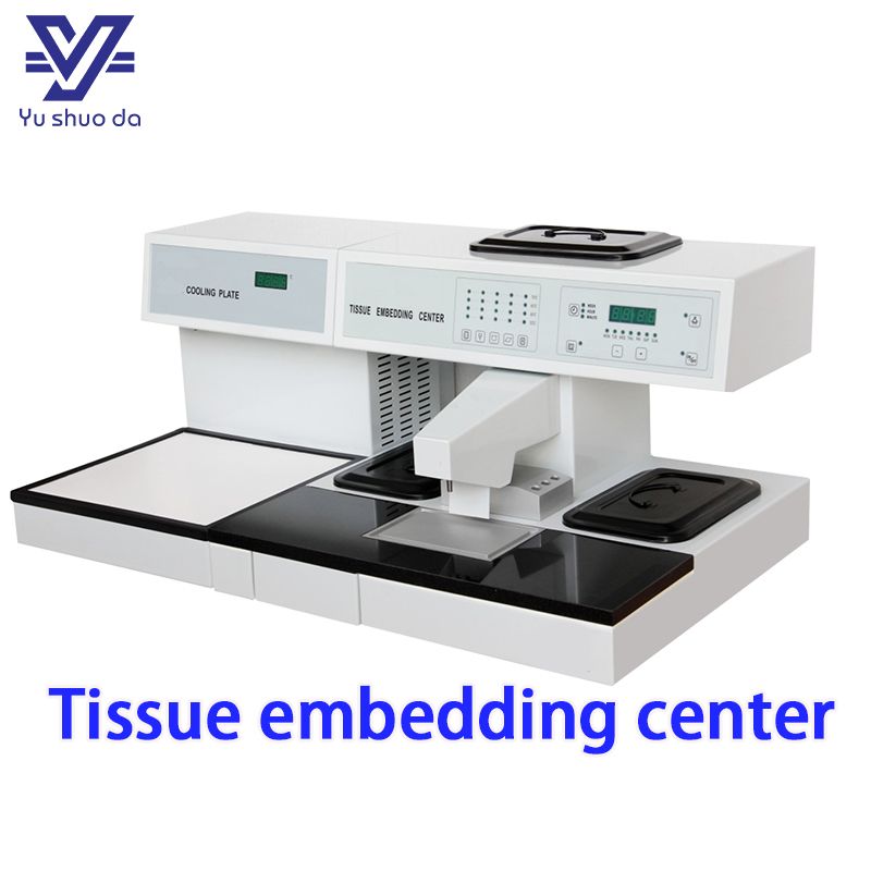
Five independently controlled heated zones plus a separate cooling module.
1. Paraffin wax running too hot. Paraffin wax for histology embedding is supplied in grades rated around 56–58 °C or 58–60 °C. Run the dispenser at 70 °C “so it flows better” and you get thin wax that holds more dissolved air and contracts harder as it sets. The voids at the block face are shrinkage cavities.
2. Air trapped during orientation. The tissue sits in an open embedding base mold and wax is poured over it. If the wax is too viscous, or poured in one fast pass, air is pinned against the tissue with nowhere to go.
3. Cooling that starts from the outside in. A block that sets on its surface first traps the last of the contraction in the centre — usually right where the tissue is.
4. Carryover from tools. Forceps and work surfaces that are not kept separate drag old wax and debris into the block.
None of these is an operator failure alone; each is a design property of the station.
The chamber and the dispensing tap do different jobs. The chamber must keep a working volume fully melted; the tap must deliver wax hot enough to flow cleanly without degrading it. One thermostat driving both means one setting is always wrong.
On a modular tissue embedding center, five heated areas work independently: the paraffin chamber, the paraffin dispenser, the left and right thermal storage compartments, and the heated working plate. Each has its own control and display, so you can hold the chamber at infiltration temperature while running the tap lower. Independent zones also remove the gradient that single-element heating creates across a large plate.
A standalone 10L paraffin dispenser with digital control from 0 to 85 °C and a 304 stainless steel inner tank gives the same precision as a separate module. The tank resists corrosion and cleans easily, and a transparent glass cover lets you watch the wax level without opening the lid. Every time you open a wax reservoir you lose heat and pull air into the melt.
Orientation is the most time-critical step in the sequence. You have to get the tissue flat, in the right plane and the right face down, before the wax beside it skins over. Cold forceps and a patchy plate shorten that window to nothing.
Look for a heated working plate with its own thermostat, and heated forceps wells that hold the tips at temperature. When forceps are not pre-warmed, wax freezes on the tips, you introduce cold spots, and the tissue shifts as you release it.
In pathology tissue embedding, the tissue embedding and cooling plate pairing decides whether the block face is flat: a tissue embedding station with a generously sized heated plate gives you time to lay the tissue down and check the plane before the wax dulls.
Two details change daily life: automatic memory and restoration of preset temperatures, so the station returns to your settings after a power interruption; and dual overheat protection, so a failed thermostat cannot cook a whole batch of wax.
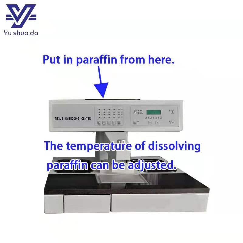
Heated forceps wells hold the orientation window open long enough to place tissue accurately.
The cooling plate is not a shelf. Fast, even, flat cooling is what prevents voids forming at the tissue face. That is why a tissue embedding center with cooling plate built as two separate modules beats a single integrated unit: you can set the cooling surface independently of wax temperature, and hold the plate at a temperature that sets the block through quickly instead of hardening the surface first.
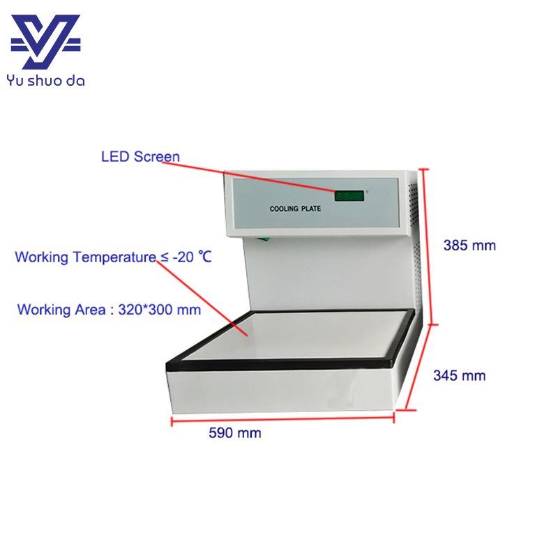
Separating the cooling module from the embedding module controls block setting independently of wax temperature.
A block set up to fail has often been failed upstream. Under-processed tissue arrives at the embedding station already shrunken, and shrunken tissue pulls away from the wax at the block face. It looks like a bubble. It is a dehydration artefact.
If you see voids and pale, shrunken tissue together, check the histology automatic tissue processor before you touch the embedding station. A carousel shape tissue processor with clean, separate reagent cups and a precisely controlled paraffin cup gives the dehydration and infiltration consistency that embedding depends on. Where dense or fibrous specimens are routine, a vacuum model tissue processor shortens the cycle and improves infiltration. The settings that matter in tissue processor histology are the same ones that matter at the block face: clean reagents, correct temperatures, enough time. Upstream of all of it, tissue has to be sampled and described properly, which starts at the grossing station.
Downstream, a slide warmer, a laboratory water bath and a slide staining jar decide whether a perfectly cut section adheres and stains evenly.
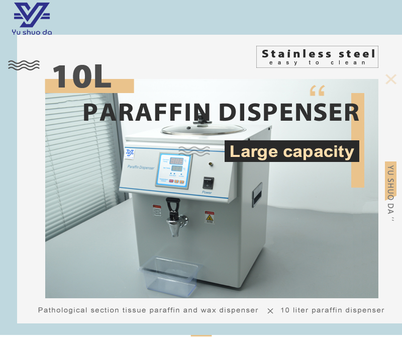
A 10L paraffin dispenser with 0–85 °C digital control and a stainless steel tank.
Whether you are specifying a biological tissue embedding center for a research laboratory, a standalone paraffin embedding center for a single bench, or an integrated pathology equipment tissue embedding center for a hospital department, the criteria are the same:
| Feature | Why it matters |
|---|---|
| Independent temperature control per heated zone | Lets the chamber and dispenser run at different, correct temperatures |
| Digital setpoint with a visible display | You can reproduce a temperature instead of guessing at it |
| Heated forceps wells | Prevents cold spots and wax build-up on the tips |
| Flat, separately controlled cooling plate | Fast, even setting without surface-first hardening |
| Automatic memory of preset temperatures | The station returns to your settings after a power interruption |
| Dual overheat protection | Protects the wax batch and the tissue if a thermostat fails |
| Low-voltage illumination | You can see the tissue face and air bubbles while you work |
Check the wax supply side too: a filter and a base-of-tank wax outlet keep sediment out of the block, and an adjustable flow valve lets you slow the pour so air is not dragged in.
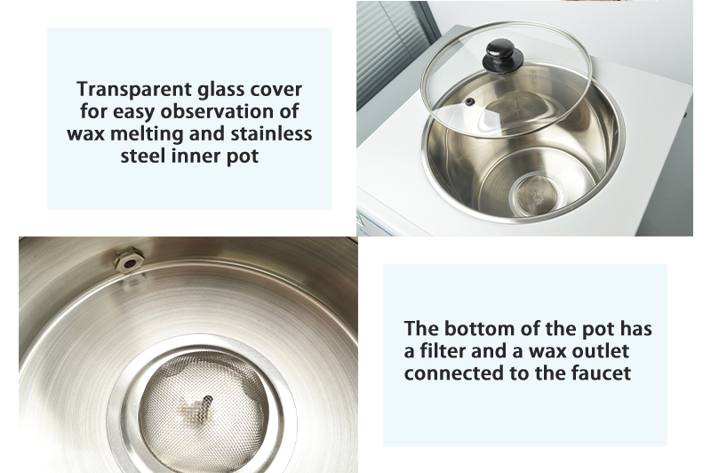
An adjustable flow valve lets you slow the pour, the simplest change that reduces trapped air.
Embedding defects often surface only later, at the rotary microtome. A block that looks perfect can still produce a poor ribbon if the face is not parallel to the blade, or if the wax is too soft to hold the tissue.
Treat the station and the microtome as one system. Set the laboratory microtome to a thickness suited to the tissue — 4 µm is the usual starting point for routine paraffin sectioning, while soft or fatty tissue may need 5–6 µm. Use a sharp tissue microtome blade and change it on a schedule, not when sections start failing: a dull pathology microtome blade compresses the block face and exaggerates every underlying void.
Choose blade geometry deliberately. A low profile microtome blade suits routine paraffin work and standard holders; a high profile microtome blade gives more rigidity for harder blocks. A disposable microtome blade removes the resharpening cycle, and pairing it with the right disposable blade holder or a dedicated microtome blade holder keeps the edge angle repeatable between operators.
For routine volume, the microtome machine needs two things: a section thickness you can set and see, and a specimen holder that does not move under load. A histology rotary microtome with a holder that advances back, forth and laterally makes trimming to the tissue face easier without re-clamping. A tissue microtome or hand microtome still has a place in teaching, but a tissue rotary microtome with a clean, measurable advance is what keeps ribbon quality stable all day.
SHENYANG YU SHUO DA SCIENCE AND TECHNOLOGY LTD (Yushuoda) has designed and manufactured histology laboratory equipment from Shenyang, China since 2016. Our range covers pathology lab equipment across the workflow: the pathology grossing station and automatic tissue processor upstream of embedding, the modular tissue embedding center and paraffin dispenser, and the rotary microtome, microtome blade holder and slide staining jar at the bench. A stainless steel workbench grossing station and a full tissue processor equipment line support laboratories scaling from a single bench to high volume, and we also supply mortuary equipment for sale, including mortuary body refrigerators and morgue refrigeration equipment. As pathology grossing station manufacturers, we build to CE certification and ship to more than 30 countries, with OEM and ODM design available.
If you are re-embedding more blocks than you should, the cause is almost always one of the four above. Send us your block size, cassette format and daily volume, and we will match a tissue embedding center configuration to your workflow. For a rotary microtome price, paraffin dispenser options, or a full embedding and sectioning quotation, send your specification and we will come back with a configuration and a lead time.
 Enviar un mensaje
Si Usted está interesado en nuestros productos y desea saber más detalles, deje un mensaje aquí, le responderemos tan pronto como nosotros ... puedamos.


 Red IPv6 admitida
 español
 English
 русский